Fetal Echocardiography
Fetal Echocardiography is a detailed and specialized ultrasound examination of the baby’s heart performed during pregnancy. It helps in early detection of congenital heart defects and allows appropriate planning of care before and after birth. Under the expertise of Dr. Ashwini Rathi, this examination is performed using advanced ultrasound technology and internationally accepted protocols.
What is Fetal Echocardiography?
Fetal Echocardiography is a targeted ultrasound that evaluates the structure, function, and blood flow of the fetal heart. Unlike routine scans, this test focuses exclusively on identifying congenital heart abnormalities in detail.
When is Fetal Echocardiography performed?
The ideal time for a fetal echocardiogram is between 18 to 24 weeks of pregnancy. However, in high-risk cases, it may be performed earlier and repeated later for confirmation.
Who should undergo Fetal Echocardiography?
- Women with diabetes, thyroid disorders, or autoimmune diseases
- History of congenital heart disease in the family
- Abnormal heart findings on routine anomaly scan
- Twin or multiple pregnancies
- Mothers with infections during pregnancy
- Exposure to certain medications or substances
- Suspected chromosomal abnormalities
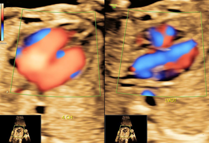
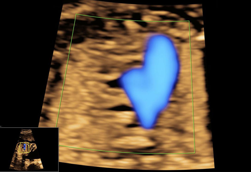
What does Fetal Echocardiography assess?
- Four-chamber view of the fetal heart
- Outflow tracts of major vessels
- Heart rhythm and rate
- Valve structure and function
- Blood flow patterns using Doppler
How is the test performed?
The procedure is performed using a high-resolution abdominal ultrasound probe. The scan is painless and non-invasive. It usually takes 30 to 45 minutes depending on the position of the baby.
You will be comfortably positioned on the examination couch, and ultrasound gel will be applied over the abdomen for optimal imaging. The fetal heart will be studied in multiple planes and angles.
Is Fetal Echocardiography safe?
Yes. Fetal Echocardiography is completely safe for both mother and baby. It uses sound waves and does not involve radiation. Dr. Ashwini Rathi follows strict international safety guidelines during the examination.
What do the results indicate?
A normal fetal echocardiogram provides strong reassurance about the baby’s heart health. If an abnormality is detected, the nature, severity, and possible outcomes will be explained in detail.
Depending on the finding, multidisciplinary counseling, follow-up scans, delivery planning at a specialized center, or postnatal intervention may be advised.
What are the limitations of Fetal Echocardiography?
Although most major heart defects can be detected, very small defects and some rhythm abnormalities may not be visible before birth. Image quality may also be influenced by fetal position, maternal body habitus, and gestational age.
Why choose Dr. Ashwini Rathi for Fetal Echocardiography?
- Extensive experience in prenatal cardiac screening
- International training and FMF-based protocols
- High-end ultrasound and Doppler technology
- Clear reporting and compassionate counseling
- Coordinated care with pediatric cardiology when required
Early detection of heart abnormalities allows timely planning and the best possible outcomes for your baby. With precise imaging and expert interpretation, Dr. Ashwini Rathi ensures thorough fetal heart evaluation and trusted prenatal care.